Pelvic Floor Insufficiency Radiology

Https Www Ajronline Org Doi Pdf 10 2214 Ajr 07 7096

Https Www Ajronline Org Doi Pdf 10 2214 Ajr 07 7096

Https Www Abdominalradiology Org Resource Resmgr Education Dfp Pelvicfloor Mri Imaging Mri Of Pelvic Floor Dysfunct Pdf

Https Www Abdominalradiology Org Resource Resmgr Education Dfp Pelvicfloor Mri Imaging Mri Of Pelvic Floor Dysfunct Pdf

Mri For Pelvic Floor Dysfunction Can The Strain Phase Be Eliminated Semantic Scholar

Mri For Pelvic Floor Dysfunction Can The Strain Phase Be Eliminated Semantic Scholar

Upright Radiology Cases

Upright Radiology Cases

Https Pubs Rsna Org Doi Pdf 10 1148 Rg 345140137

Https Pubs Rsna Org Doi Pdf 10 1148 Rg 345140137

Figure 3 22 From Mri Of Pelvic Floor Dysfunction Review Semantic Scholar

Figure 3 22 From Mri Of Pelvic Floor Dysfunction Review Semantic Scholar

Figure 3 22 From Mri Of Pelvic Floor Dysfunction Review Semantic Scholar

Pelvic floor failure is a common disorder that affects 23 7 of women in the united states with a prevalence of 9 7 49 7 that increases with age one in nine women will undergo an invasive procedure for treatment of urinary incontinence or pelvic organ prolapse with 30 requiring additional surgery for symptom recurrence by 80 years of age.

Pelvic floor insufficiency radiology.

In this article we focus on imaging pelvic vein insufficiency and related extending varicosities. Pelvic congestion syndrome pcs is a chronic condition that occurs in women when varicose veins form below the abdomen within the pelvic region. Pelvic venous congestion is a commonly overlooked condition that can be severely painful and debilitating for many women. Since pelvic floor imaging is a dynamic form of imaging aiming to evaluate the evacuation phase rectal filling is mandatory.

223 501 508 google scholar 17. Varicose veins are veins that become swollen. The diagnosis of pelvic venous insufficiency is therefore essentially a clinical exercise that is supported by radiology findings. Pelvic venous congestion syndrome is also known as ovarian vein reflux.

It is a cause of chronic pelvic pain in approximately 13 40 of women. Authors have used different filling media such as sonographic gel mashed potatoes variably mixed with a gadolinium compound according to the signal intensity intended to obtain in the t1 or t2 weighted sequences. Fielding jr griffiths dj versi e mulkern rv tee mlt jolesz fa. Mr imaging of pelvic floor continence mechanisms in the supine and sitting positions.

The term pelvic congestion syndrome specifically refers to the condition first described by louis alfred richet in 1857 and characterized by chronic dull pelvic pain pressure and heaviness that persist for more than 6 months with no other cause. Pelvic venous insufficiency pvi is a condition resulting from broken valves on the inside of the gonadal ovarian veins. Venography is primarily carried out for treatment purposes before embolization. That extend from the leg through the pelvic floor which should be evaluated for the presence of pelvic congestion syndrome.

The disease process was previously referred to as pelvic congestion syndrome however the term pelvic venous insufficiency identifies the root cause and is the newer terminology. The imaging examinations also allow other causes of cpp to be excluded. Pelvic floor dysfunction is the inability to control the muscles of your pelvic floor. Chronic pelvic pain is pain in the lower abdomen which has been present for more than 6 months.

Dynamic mr imaging of the pelvic floor performed with patient sitting in an open magnet unit versus with patient supine in a closed magnet unit.

Pelvic Floor Lipoma Radiology Case Radiopaedia Org

Pelvic Floor Lipoma Radiology Case Radiopaedia Org

Translabial Us And Dynamic Mr Imaging Of The Pelvic Floor Normal Anatomy And Dysfunction Radiographics

Translabial Us And Dynamic Mr Imaging Of The Pelvic Floor Normal Anatomy And Dysfunction Radiographics

Https Pubs Rsna Org Doi Pdf 10 1148 Rg 284075139

Https Pubs Rsna Org Doi Pdf 10 1148 Rg 284075139

Magnetic Resonance Imaging Of The Pelvic Floor Sciencedirect

Magnetic Resonance Imaging Of The Pelvic Floor Sciencedirect

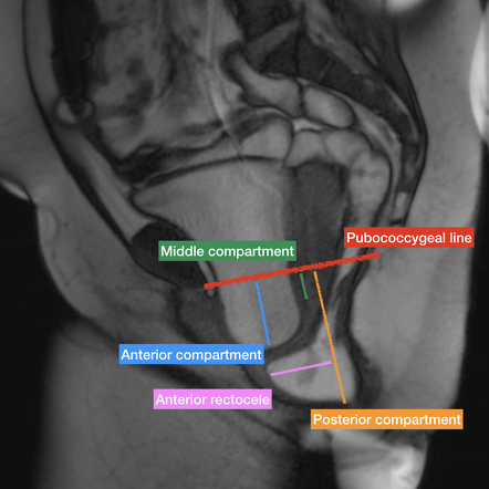
Pubococcygeal Line Radiology Reference Article Radiopaedia Org

Pubococcygeal Line Radiology Reference Article Radiopaedia Org

Can We Correlate Pelvic Floor Dysfunction Severity On Mr Defecography With Patient Reported Symptom Severity Springerlink

Can We Correlate Pelvic Floor Dysfunction Severity On Mr Defecography With Patient Reported Symptom Severity Springerlink

Ic And Pelvic Floor Dysfunction Treat Ic

Ic And Pelvic Floor Dysfunction Treat Ic

Dynamic Magnetic Resonance Imaging Of The Female Pelvic Floor A Pictorial Review Springerlink

Dynamic Magnetic Resonance Imaging Of The Female Pelvic Floor A Pictorial Review Springerlink

Physiotherapy First For Pelvic Floor Dysfunction Urology News

Physiotherapy First For Pelvic Floor Dysfunction Urology News

Joint Esur Esgar Recommendationsfor Reporting Of Pelvic Floor Dysfunction Pfd European Radiology

Joint Esur Esgar Recommendationsfor Reporting Of Pelvic Floor Dysfunction Pfd European Radiology

How To Perform And Report Magnetic Resonance Imaging For Pelvic Floor Dysfunction An Interactive Case Based Approach Springerlink

How To Perform And Report Magnetic Resonance Imaging For Pelvic Floor Dysfunction An Interactive Case Based Approach Springerlink

Dry Needling In Myofascial Tracks In Non Relaxing Pelvic Floor Dysfunction A Case Study Sciencedirect

Dry Needling In Myofascial Tracks In Non Relaxing Pelvic Floor Dysfunction A Case Study Sciencedirect

Https Www Abdominalradiology Org Resource Resmgr Education Dfp Pelvicfloor Anatomy Pelvic Floor Imaging And Ana Pdf

Https Www Abdominalradiology Org Resource Resmgr Education Dfp Pelvicfloor Anatomy Pelvic Floor Imaging And Ana Pdf

Dynamic Mr Imaging Of The Pelvic Floor A Pictorial Review Radiographics

Dynamic Mr Imaging Of The Pelvic Floor A Pictorial Review Radiographics

Pelvic Floor Imaging Radiology

Pelvic Floor Imaging Radiology

Epos Trade

Epos Trade

Academic Onefile Document Role Of Dynamic Mri In Assessment Of Pelvic Floor Dysfunction In Females

Academic Onefile Document Role Of Dynamic Mri In Assessment Of Pelvic Floor Dysfunction In Females

Evidence Based Prevention And Treatment Of Pelvic Floor Dysfunction

Evidence Based Prevention And Treatment Of Pelvic Floor Dysfunction

Https Encrypted Tbn0 Gstatic Com Images Q Tbn 3aand9gcqizonqboo3beyyikracw6diewfrutuxrrlwy Vesvyqdb 7njk Usqp Cau

Https Encrypted Tbn0 Gstatic Com Images Q Tbn 3aand9gcqizonqboo3beyyikracw6diewfrutuxrrlwy Vesvyqdb 7njk Usqp Cau

Dynamic Imaging Of Posterior Compartment Pelvic Floor Dysfunction By Evacuation Proctography Techniques Indications Results And Limitations European Journal Of Radiology

Dynamic Imaging Of Posterior Compartment Pelvic Floor Dysfunction By Evacuation Proctography Techniques Indications Results And Limitations European Journal Of Radiology

Figure 14 Magnetic Resonance Imaging Of Female Pelvic Floor Dysfunction A Review Of Dynamic Mri Defecography Springerlink

Figure 14 Magnetic Resonance Imaging Of Female Pelvic Floor Dysfunction A Review Of Dynamic Mri Defecography Springerlink

Pdf Pelvic Floor Ultrasound

Pdf Pelvic Floor Ultrasound

Pelvic Floor Disorders Pelvic Girdle Pain And Symphysis Pubis Dysfunction Following Sports Injury Caring Medical

Pelvic Floor Disorders Pelvic Girdle Pain And Symphysis Pubis Dysfunction Following Sports Injury Caring Medical

X Ray Of The Pelvis With A Pubic Symphysis Separation Is Shown In A Download Scientific Diagram

X Ray Of The Pelvis With A Pubic Symphysis Separation Is Shown In A Download Scientific Diagram

Source : pinterest.com